Are scanners safe at airports. Scanners at the airport
For flight safety, scanners are installed at each airport. Basically, they are divided into two types - metal detectors (the so-called frames) and scanners that create a complete image of the human body on the screen. The latter are considered more effective for searching for items prohibited from being carried on board the aircraft.
Unlike the frame of a metal detector, passing through which a person does not leave any "traces", passing through the scanner creates an image very similar to a naked body. Airport security services claim that the anonymity of air passengers is fully respected - the scanner operator does not see the face, the scanned images are not saved.
Technologies
Human body scanners are based on two different principles. The first of them is similar to an X-ray machine, but the rays do not pass through, but are reflected. Materials of different density are displayed in different colors. Less dense - skin, muscles - light, and more dense metal - dark. For getting detailed information on such a scanner, you need to take two pictures: front and back. The full name of this scanner is x-ray scanner based on backscatter (Backscatter X-ray).

The second full-sized scanner uses millimeter waves in its work, emitted by two antennas rotating around a person. Clothing and other similar materials are transparent to the radiation of the scanner, and the image is very clear and detailed, much more realistic than from an X-ray scanner.


How to distinguish
It is very easy to distinguish between scanners. The magnetic frame - the metal detector looks like the letter "P", through which you have to pass, and in the body scanners you have to stop. An X-ray scanner is like two booths, between which a passenger stands, and a microwave scanner is a small transparent booth where they pass around the antenna once.
Safety and health
Manufacturers claim that both types of scanners are completely safe. A microwave scanner can be compared to the radiation from a mobile phone, and an X-ray gives the same exposure as 2 minutes of flight on an airplane. Despite such assurances, an X-ray scanner is used much less frequently at airports, and children and pregnant women are not taken through it. Wherein big studies health safety checks for microwave and X-ray scanners have not been conducted. Latest publications from scientists different countries express concern about the possible influence of scanners on the formation of tumors and mutations in DNA. But there is no documentary evidence.
The second big problem with scanners is the reluctance of passengers to show themselves in an unsightly way, even to an anonymous operator. It is technically possible for programs that serve scanners to save the received images, but all countries that have introduced such technologies have promised not to use this function. Unfortunately, it turned out that images from airports are saved on the computers of operators, and leaks of such images have already appeared.
Alternative
If you don't want to go through the detailed image scanner, you can opt out. Since it is not possible to fly without following certain security screening procedures, most airports will offer manual screening instead of scanning. The screening is carried out by a security officer of the same gender as the passenger and does not require privacy in the booth or undressing. Screening technology differs from country to country.
In the United States, until recently, the regulations provided for manual inspection with the back of the hand, but recent instructions have changed the rules - now the palpation is carried out with the fingers. If you refuse screening at US airports, unpredictable problems are possible, up to calling the police.
At Domodedovo Airport, a manual screening is no different from an additional screening after a metal detector has gone off, and refusing to pass through the scanner does not cause any problems or additional questions.
In the event that manual screening is unacceptable for one reason or another, there is only one option left - to refuse to fly through a specific airport where the scanner is installed.
List of airports
In Russia, a new type of scanner has been installed at Domodedovo airport for about 3 years. This is the L3 Provision microwave scanner. Similar ones are installed at Sheremetyevo, but they are not always used and passengers are escorted through a more familiar frame.
Most scanners are installed in the USA. There are now almost 500 scanners in US airports, with 500 more to come during 2011. At the airports of New York (JFK), Chicago (ORD) there are scanners using X-rays, in Miami, Las Vegas and Los Angeles - microwave ones.
In Europe, scanners are installed in Amsterdam, London (LHR), Hamburg, Manchester.
From almost complete list the type of scanner and its location can be found on the FlyerTalks website -
Doctors are sounding the alarm - after the recent terrorist attacks at airports, they want to install x-ray scanners that can cause cancer. There is talk of installing devices for covert screening even in the Moscow metro. Is it worth it to refuse to pass through when you next fly to rest on the sea and is it possible to do this?
At the beginning of March 2011, Rospotrebnadzor issued an official statement: installations for X-ray scanning of people at airports are dangerous. Chief Sanitary Doctor of the Russian Federation Gennady Onishchenko believes that X-ray scanners can lead to radiation and oncological diseases.
Modern research confirm the fears of doctors. UC San Francisco biochemist and biophysicist David Agard argues that any dose of x-ray radiation, even the smallest, is harmful. He believes that X-rays cause chromosomal rearrangements that provoke the growth of cancer cells. The installation of such devices at airports will lead to an increase in the incidence of melanoma and breast cancer.
Whoat risk?
X-rays are most dangerous for pregnant women. If exposed to radiation in the womb, in the future it threatens the risk of developing cancerous tumors and leukemia. David Brenner, head of the Center for Radiological Research at Columbia University, adds that 5% of the population is genetically sensitive to radiation. These people have an increased risk of developing basal cell skin cancer.
Separately, it should be said about people who, on duty, are forced to constantly be in the air. Pilots and flight attendants go through x-rays about 400 times a year. These figures for "business travelers": journalists, businessmen and diplomats are about two times lower. Add the cosmic radiation that passengers are exposed to while flying at a cruising altitude of 10 km. The farther a person is from the surface of the planet, the more he is irradiated.
In fact, we can all be at risk at any moment: sophisticated equipment can simply fail, and then any passenger will receive a serious dose of radiation. Yes, and serviceable is a dangerous thing: manufacturers underestimate dangerous indicators in order to obtain a safety certificate.

How it works?
X-ray operates on the principle of backscattering, when two extremely weak X-ray beams form a two-dimensional image on the screen. The Food and Drug Administration (USA) assures that the devices are absolutely safe for all passengers, including the elderly, children and people with implants. According to the specialists of the Office, a person needs to go through more than 1000 times a year, and only after that it can be said that the exposure standards have been exceeded.
There is a safer alternative to the X-ray scanner, the microwave. The car directs a beam at the passenger and then analyzes the reflected signal. However, the installation sparked many protests. The device allegedly shines through clothing, allowing literally"Look under your skirt"
What to do?
At Sheremetyevo and Vnukovo, X-ray scanners are used only for baggage screening. Passengers pass through rapiscans, the radiation of which is 1000 times weaker than radiation mobile phones.
However, you can choose not to pass through any device. The security officer will then conduct a search in the security room. In this case, the passenger will be searched by a woman, and the passenger by a man.
Oleg Polyakov, Deputy Chief Physician of the Russian Medical Academy of Postgraduate Education, comments on the situation: “X-ray is a serious examination. For each patient, his own dose is calculated, taking into account the weight and age of the patient, the location of the organ that needs to be examined. All radiation is recorded, the annual dose of radiation is calculated. It is hard to imagine what will happen to people who are forced to fly frequently for work. But there are still people with thyroid diseases, oncology, those who have recently done fluorography. Irradiation does not go away on its own, it accumulates. If X-ray machines are installed on vehicles, who will monitor the radiation doses for each individual?”
Numbers
- The maximum permissible background for life is 5 mSv per year (millisievert)
- Tooth snapshot - 1 µSv
- Inspection using the SibScan microdose X-ray system installed at Pulkovo and Domodedovo airports - 0.5 μSv
- Irradiation during the flight Moscow - Bangkok - 45 μSv
- Exposure during the flight Moscow - Sharm el-Sheikh - 30 μSv
- Exposure during the flight from Singapore to New York - 90 μSv
- Film fluorography (outdated technology, being replaced by digital) - 500-800 µSv
- Digital fluorography - 60 μSv
- The lethal dose of radiation is just under 1 sievert.
At airports around the world, X-ray backscatter scanners are used to screen passengers and baggage ( English Backscatter X-ray scanner). This is the same apparatus that at one time caused a number of scandals due to the fact that it “undresses” people.
American do-it-yourselfer Ben Krasnow assembled a working model of such a scanner from parts purchased on ebay (author's description). Here is an example of an image taken by Ben:
Did you find out what it is?
That's right, it's a turkey in a Christmas sweater: 
In addition, she tried to carry a hex key inside herself, which was easily detected by the scanner. 
Principle of operation
Unlike first-generation screening systems and medical X-ray machines, backscatter scanners detect radiation that has not passed through an object, but reflected from it. Backscattering of X-rays is primarily due to the Compton effect. While transmission scanners make it possible to obtain only the distribution of the density of a substance, backscattering devices are capable of distinguishing the composition of a material, including an organic one.The scanner consists of an x-ray tube with a scanner (in the photo - in the middle), an x-ray detector (left) and power supplies (right). 
Radiation source
The source of X-rays is this tube:
It works as follows: electrons emitted by the cathode (right) are accelerated by a strong electric field and enter the massive anode (left). Upon sudden deceleration in the anode material, the electrons generate X-rays. Due to the beveled surface of the anode, the radiation is reflected to the side and leaves the tube. To power the tube, a high-voltage source of several tens of kilovolts is needed.
The tube is placed in a metal case with a narrow exit slit. Opposite the slit there is a collimator disk with a small hole, which makes a thin beam from a wide beam of radiation. 
During the operation of the installation, the disk is rotated by the engine, and the beam moves horizontally, drawing line by line. Vertical scanning is carried out by turning the tube along with the body and disk around the horizontal axis. While this is done manually, but the design provides for the installation of a second motor.
Detector
The detector consists of a luminescent screen and a photomultiplier tube (PMT) in an opaque housing.
Under the action of X-rays scattered by the object, the screen begins to glow. The PMT converts this light into an electrical signal. The signal from the PMT is amplified by a simple amplifier and fed to the oscilloscope, to the brightness control input (Z-input). 
The horizontal sweep of the oscilloscope is synchronized with the rotation of the collimator, so one line of the image is visible on the oscilloscope. 
The device for vertical deflection of the beam is equipped with a potentiometer, the signal from which is fed to the Y-input of the oscilloscope. Thus, when the X-ray beam is deflected up and down, the line on the oscilloscope screen moves accordingly. To compose a complete picture from individual lines, Ben simply photographs the oscilloscope screen with a long exposure. 
The image is not very clear, noisy, but the contours of the object and contrast elements are clearly guessed in it. internal structure(for example, a key in a turkey).
Video
Ben talks about his setup and demonstrates it in action:The story about the X-ray detector and photomultiplier:
X-ray scanners- Devices used to obtain fluoroscopic images. These scanners are used in various areas: security, flaw detection, etc. Sometimes medical x-ray machines are called x-ray scanners.
Encyclopedic YouTube
1 / 1
Warehouse errors
Subtitles
Story
The first X-ray image was obtained from the hand of V. Roentgen's wife. The image showed her wedding ring on her finger, as well as the bones of her hand. January 18, 1896 "X-ray machine" was officially presented by H. Smith, new car was presented to the public as a technical miracle and was intended mainly [ ] for fun. Circus performers used these devices to show the public their skeletons and hand out X-ray images of hands with jewelry on their fingers. While many people were fascinated by the discovery of such devices, some were concerned about the possibility of using such devices to look through doors and violate privacy.
In the 1940s, 50s X-ray scanners were used in stores to help sell shoes (the picture showed how well the shoes fit the customer). From the moment when the harmful effect of X-rays was discovered, the use of such scanners almost instantly ceased.
Review
An X-ray scanner usually consists of an X-ray source (accelerator or X-ray tube) and a detection system, which can be in the form of a film (analogue technology) or a detector line or array (digital technology).
luggage scanners
X-ray scanners are used for non-contact screening of cargo and luggage for the possible presence of weapons, drugs and explosives. X-ray radiation is localized inside the body of the scanners and therefore they are safe for others. The main part of such scanners is an X-ray generator, a detector line for detecting rays passing through the checked baggage, a data processing unit for converting the signals received from the detector line into an image, and a conveyor that is used to guide the baggage through the scanner. The resulting images are displayed on a computer terminal, usually located near the scanner.
Purpose:
In modern conditions, only the use of electromagnetic metal detectors for screening a person and X-ray systems for screening luggage no longer provides the required level of security and control. First of all, this concerns security at airports and customs control at the border, when cold non-metal weapons, plastic explosives are used to commit terrorist acts, drugs are illegally transported in swallowed capsules.
In addition, the task of increasing the level of security is important:
- on the border with states in which armed conflicts occur,
- at protected sites
- VIP persons,
- in mines and enterprises associated with the extraction and processing of diamonds, precious stones and metals, rare earth elements.
The main problem is that the weapons of terrorism and objects prohibited for transportation can be made of non-metallic materials and be hidden not only under clothing, but also in natural body cavities. To date, digital human scanning systems based on the use of X-ray radiation are becoming a fundamentally new means of control.
Scanning digital radiography technology for safety:
The unique technology for obtaining a full-length digital projection X-ray image of a person using the “flat beam scanning” method is based on:
- on the use of an ultra-highly sensitive linear array of semiconductor scintillation detectors located vertically as an X-ray receiver;
- on the formation of an extremely narrow (less than 2 mm) monochromatic X-ray beam using a system of collimators and filters in order to minimize the exposure of a controlled person;
- on the movement of a person on a special moving platform located between the collimator and the detector through the X-ray beam, for the purpose of scanning and personal inspection;
- on the detection of radiation that has passed through a person using a linear detector and the formation of a two-dimensional matrix of a digital image on the operator's monitor;
- on optimizing the ratio of radiation dose and resolution, depending on the specifics of the problem being solved.
A digital X-ray scanner using this technology allows you to get a projection image of a controlled person and actually makes it possible to “look inside” a person for the purpose of personal inspection. At the same time, there is no obvious ethical problem, which is typical for scanning X-ray systems based on reflected radiation, which, as it were, "undress a person." In addition, two scans are not required - in front, behind and in some cases from the side to screen a person.
The X-ray scanner is designed to detect dangerous objects:
- from inorganic materials hidden under clothing - firearms and cold steel, fuses, electronic devices, etc.;
- from organic materials (materials not detected by a metal detector) hidden under clothing - plastic explosives, drugs in containers, ceramic firearms and cold weapons, etc.;
- from materials of any type, swallowed or hidden in natural cavities of a person - drugs, explosives, chemical and biological substances in containers, precious stones and metals.
Main applications:
- at airports, railway and bus stations to ensure the safety of mass passenger traffic;
- at protected facilities for the purpose of input / output control;
- in prisons as an alternative to strip searching.
Additional applications:
- at the border for the purpose of customs inspection to detect contraband;
- in mines and factories for the extraction of diamonds in order to prevent theft;
- in workshops for finishing and processing of diamonds, precious stones and metals, concentrates of rare earth elements in order to prevent theft;
- VIP security.
Installations:
Currently SecureScan systems are installed and operated by:
- INCHEON airport ( South Korea) - 2 systems
- Diamond mines ( South Africa, Angola) - 5 systems
- Prison in Lodz (Poland) - 1 system
- Los Angeles Prison (USA) - 1 system
- Security VIP ( Saudi Arabia) - 1 system
- Airport (Turkey) - 1 system.
The SecureScan system was tested in airports in Orly(France), Amsterdam (Netherlands), UAE, Qatar, etc.
Safety:
Human exposure dose per scan:
- in ultra-low dose mode does not exceed 0.1 μSv;
- in high resolution mode does not exceed 3-5 μSv.
In terms of radiation safety, the system complies with the American National Standard ANSI/HPS N43.17-2002 “Radiation Safety For Personnel Security Screening Systems Using X-rays”.
Security for the person being scanned:
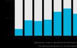
For comparison, the typical dose due to cosmic radiation for a person making a one-way flight from Malaga to London is 10 µSv, from New York to London 35 µSv and from Hong Kong to London 50 µSv. Typical background radiation dose received by the average representative of countries European Union per day is 6-7 μSv.
Thus, the exposure to which a person is exposed on a scanning x-ray system, slightly against the background of natural radiation. Being in the sun or any flight on an airplane contributes dozens of times more to the total human exposure.
According to the recommendations of the American national council according to radiation protection (NCRP 1993) and international safety standards for the general contingent of the population (pregnant women and children among them), an exposure level of 1 mSv (1000 μSv) per year from all sources of ionizing radiation for non-medical purposes is acceptable. If we take as a basis a value four times smaller - 0.25 mSv, then on this system even pregnant women and children without harm to health can be subjected to up to 2500 scans per year, which is obviously unrealistic for reasons of common sense.
When using the scanner on protected objects with a pass (using special cards) access system (for example, on diamond mines and factories), it is not difficult to organize the control of personal accumulated dose. In this case, even when the system is in the high resolution, total number examinations on the system can be more than 300 times a year and inspection during the year can be organized in such a way as not to allow the accumulated dose to be exceeded. Objectively, this is quite enough to ensure security, given the presence of strict control and a limited number of visits to any protected facility.
Safety for the operator:
The system can be controlled by one or several operators. If the operator is within the working area at a distance of less than 1.5 m from the scanner, his protection at the workplace is provided with a special protective screen made of lead glass, which allows you to observe the controlled person. The dose received by the operator outside the work area does not exceed the typical background dose and no additional X-ray protection is required.
Safety for others:
Outside the working area at a distance of more than 1.5 m from the scanner, the level of X-ray radiation does not exceed the background value and therefore does not pose any danger to others. This allows you to place the scanning system in a compact area in crowded places, for example, at airports next to the baggage screening system.
Specifications:
| Characteristics of the digital image | |
| Digital Image Size (Scan Margins) | 2000x800mm (ultra low dose); 2000x800 mm (high resolution) |
| Image matrix format, pixels | 672 x 275 (ultra low dose); 2688x1100 (high resolution) |
| Spatial resolution | |
| low contrast objects | 5-7 mm (ultra-low dose); 1-2 mm (high resolution) |
| high contrast objects | 0.3 mm (ultra low dose); 0.2 mm (high resolution) |
| Scan time | 8 sec (ultra low dose); 16 sec (high definition) |
| Average viewing time | 5 (ultra low dose); 10 (high definition) |
| physical characteristics | |
| Dimensions | 2100 x 4500 x 2400 mm |
| Weight | 1300 kg |
| Network Requirements | 220/110V, 50/60Hz |
| Power consumption | no more than 6 kW |
| Characteristics of the x-ray generator: | |
| Working anode voltage | 160 kV |
| Working anode current | 2.5 mA |